Endoscopes have changed how clinicians investigate the digestive, respiratory, and urinary systems. Instead of relying only on scans or symptoms, doctors can view internal tissue directly through a small flexible instrument. They may see a bleeding vessel, a shallow ulcer, or an irregular patch that imaging could miss. This visual evidence often guides faster, more focused decisions.
So, how do endoscopes improve diagnostic accuracy? High-definition cameras reveal fine surface changes, while enhanced imaging modes can emphasize abnormal blood vessels and tissue patterns. A clinician can also collect a biopsy during the same procedure, sending suspicious tissue to a laboratory for microscopic examination. This combination of direct observation and pathology creates stronger evidence than either method alone. Real-time documentation also helps specialists review findings, compare images, and monitor changes during follow-up care.
Still, endoscopy is not infallible. Small lesions may hide in folds, and inflammation can resemble more serious disease. Image quality depends on preparation, equipment maintenance, and the examiner’s experience. An uncomfortable patient may move, limiting the view. These details matter. Professional guidelines, careful training, complete reporting, and appropriate biopsy decisions help reduce errors. However, clinicians must interpret endoscopic findings alongside medical history, laboratory results, imaging, and pathology. A clear picture is valuable, but it is not always a complete answer. Understanding these strengths and limitations explains why endoscopes remain powerful tools for reliable diagnosis.
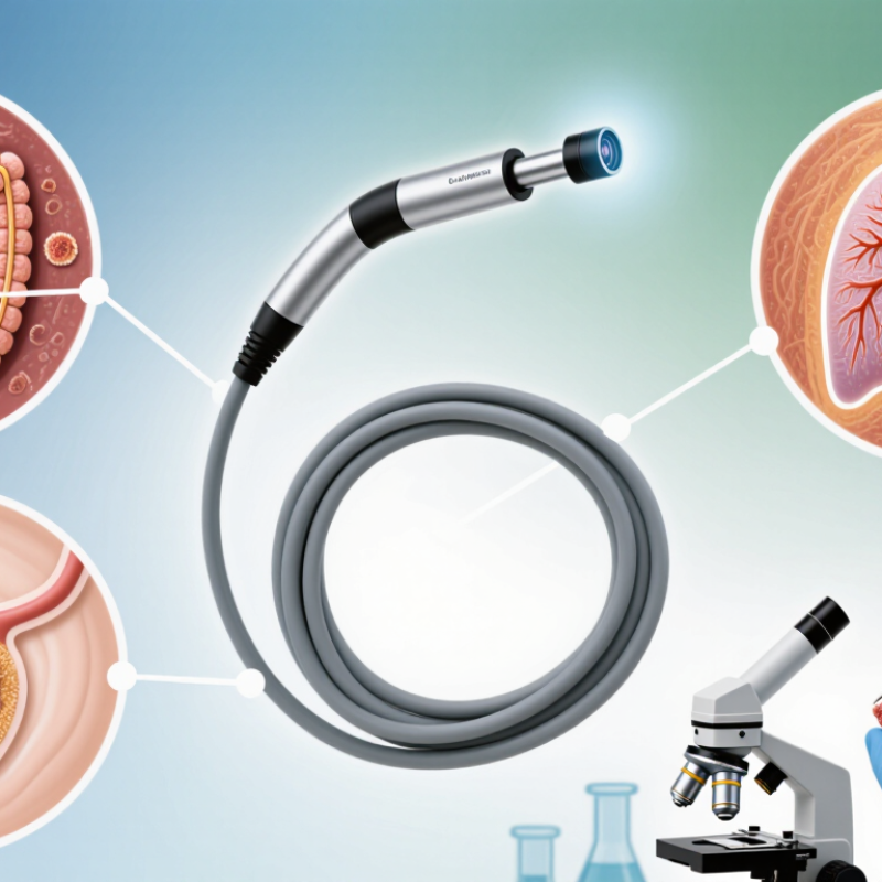
Diagnostic endoscopes are selected according to the body area and the question a clinician needs to answer. A flexible upper endoscope passes through the mouth to examine the esophagus, stomach, and upper small intestine. Its camera can reveal redness, ulcers, or narrowing. Small tissue samples may help clarify what an image alone cannot.
A colonoscope examines the large intestine, often helping clinicians identify polyps, inflammation, or bleeding. A bronchoscope travels into the airways to inspect the lungs and collect fluid or tissue when needed. Cystoscopes examine the bladder through the urethra. These instruments differ in length, shape, and working channels, so one type cannot simply replace another. Fit matters.
Some examinations use rigid endoscopes, especially when a straight, stable viewing path is useful, such as in certain joint procedures. Capsule endoscopy offers another approach: a swallowed camera records images as it moves through the digestive tract. It can show areas that standard scopes may not reach easily, but it cannot take a biopsy or treat a finding. No scope sees everything. Image quality, preparation, anatomy, and operator judgment all affect what can be detected. Even a clear picture may leave questions, which is why findings are interpreted alongside symptoms and other tests.
| Endoscope Type or Technique | Common Diagnostic Examination | How It Can Improve Diagnostic Accuracy | Typical Findings or Targets | Important Considerations |
|---|---|---|---|---|
| High-definition white-light endoscope | Upper endoscopy and colonoscopy | Provides a detailed view of the gastrointestinal lining, helping clinicians identify visible abnormalities and select areas for closer inspection or biopsy. | Ulcers, inflammation, polyps, bleeding sources, and visible tumors | Image quality depends on adequate preparation, visibility, careful inspection, and operator experience. Subtle or flat lesions may be difficult to detect. |
| Narrow-band imaging (NBI) endoscope | Upper endoscopy and colonoscopy with enhanced optical imaging | Uses filtered light to emphasize superficial blood vessels and mucosal patterns, which can help characterize selected lesions and guide targeted examination. | Abnormal vascular or surface patterns in selected esophageal, gastric, and colorectal lesions | It complements, rather than replaces, standard white-light inspection and histopathology. Performance varies with lesion type and image quality. |
| Chromoendoscopy | Colonoscopy or upper endoscopy using topical contrast dyes | Highlights differences in surface texture and lesion borders, helping direct attention to subtle mucosal changes and guide targeted biopsy. | Flat or subtle lesions, including areas of abnormal mucosal pattern | Requires careful application and interpretation. Dye-based examination may take additional time and does not by itself confirm a diagnosis. |
| Endoscopic ultrasound (EUS) | Examination of the digestive tract and nearby structures | Combines endoscopy with ultrasound to show structures beneath the lining and nearby organs, supporting assessment of lesion depth and regional features. | Pancreatic lesions, bile-duct abnormalities, and gastrointestinal wall or nearby lymph-node findings | Findings may require confirmation with tissue sampling. Results depend on the indication, image quality, and specialist interpretation. |
| Capsule endoscope | Small-bowel examination | Captures images as a swallowed capsule passes through the digestive tract, allowing visualization of much of the small bowel that may be difficult to reach with standard scopes. | Small-bowel bleeding, mucosal lesions, and findings associated with suspected small-bowel disease | It cannot take biopsies or perform treatment. In selected patients, capsule retention is possible, particularly if narrowing is present. |
| Confocal laser endomicroscopy (CLE) | Selected advanced endoscopic examinations | Provides magnified, real-time microscopic images of tissue at the examination site, which may help guide targeted sampling in appropriate settings. | Microscopic features of selected mucosal abnormalities | It is an adjunct technique, not a substitute for standard histological examination. Availability and use vary by clinical setting. |
| Endoscopic biopsy | Upper endoscopy, colonoscopy, and other endoscopic procedures | Collects tissue from a visually identified area for laboratory examination, helping confirm or clarify diagnoses that cannot be established from appearance alone. | Suspected inflammation, infection, precancerous changes, or malignancy | Sampling is limited to the tissue obtained. The result should be interpreted alongside the endoscopic appearance and the patient’s clinical context. |
A small camera captures tissue color, surface patterns, and shape. Flexible instruments can show curved passages from different angles.
Images may show tiny vessels, shallow ulcers, or irregular tissue edges. Small details can matter.
Distance, focus, lens cleanliness, lighting, and instrument movement all matter. Mucus or blood can also block the view.
They may rinse the area, adjust the angle, or reposition the instrument. Slow, careful movement can expose hidden surfaces.
Yes. Clinicians can pause and inspect subtle color changes, uneven surfaces, or bleeding as they appear.
No. Motion blur, glare, narrow spaces, or folds may hide details. A clear screen is not a guarantee.
Clinicians can mark suspicious areas and compare views from later visits. This makes changes easier to review.
A clinician can collect tissue while an area is visible. Laboratory testing can add information that images alone cannot provide.
No. Images support judgment, but interpretation depends on training and examination conditions. Uncertainty remains.
Endoscopes improve diagnostic accuracy by allowing clinicians to examine internal structures directly, using instruments suited to different parts of the body. Flexible endoscopes can navigate curved passages, while rigid designs may be used where a stable, straight view is appropriate. Their illumination and imaging systems capture detailed views of tissue, helping clinicians assess surface changes that may be difficult to detect through external examination alone. Understanding how do endoscopes improve diagnostic accuracy begins with this combination of targeted access, clear imaging, and careful observation.
Real-time visualization lets clinicians inspect suspicious areas, compare nearby tissue, and guide further examination as it happens. When needed, an endoscope can also help collect small tissue samples for laboratory analysis, adding evidence that visual inspection alone cannot provide. Diagnostic accuracy depends on image quality, the clinician’s experience, patient preparation, the instrument’s suitability, and whether the area of concern is fully visible. Together, these factors help support a more informed diagnosis while reducing reliance on indirect signs alone.

 TC MediGroup