How to Integrate China Top Imaging Systems in Surgery?

Time:2026-10-17 Author:Oliver
0%

Integrating China’s top imaging systems into surgery requires more than purchasing advanced hardware. It requires clinical planning, technical testing, and responsible team training. The practical question is how to integrate imaging systems in surgical operations without interrupting sterile workflows or delaying critical decisions. Surgeons may need real-time ultrasound, cone-beam CT, endoscopy, or navigation images beside the operating table. Each system should connect reliably with surgical displays, hospital networks, and electronic medical records. DICOM compatibility matters, but it does not guarantee smooth performance. Latency, image calibration, room layout, and backup procedures also influence patient safety.

The strongest integration plans begin with a defined clinical purpose. A spine team may prioritize navigation accuracy, while an oncology unit may need rapid image fusion. Experienced engineers should test data transfer before live procedures. Clinicians should verify image quality using realistic cases, not only laboratory demonstrations. Cybersecurity controls, user access rules, and software updates deserve equal attention. Local regulatory requirements must guide procurement, validation, and maintenance. China-based manufacturers may offer competitive imaging performance, responsive service, and adaptable configurations. Still, hospitals should examine independent evidence, warranty terms, training quality, and long-term support. No integration plan is perfect. Small workflow problems often appear only after repeated use. Regular audits, staff feedback, and documented corrective actions can turn those weaknesses into safer, more dependable surgical practice.
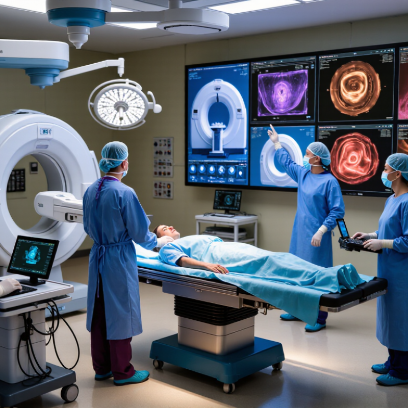
How to Integrate China Top Imaging Systems in Surgery?

Understanding China’s Top Imaging Systems for Surgical Use

China’s top imaging systems for surgical use should be judged by clinical performance, integration, and service reliability.

A high-resolution C-arm or intraoperative CT can support navigation, fracture fixation, and tumor procedures. However, image quality alone is not enough. The system should connect smoothly with PACS, hospital information systems, and DICOM workflows.

Grand View Research estimated the global medical imaging market at more than USD 40 billion in 2023. This growth reflects rising demand for faster and more precise surgical imaging. China’s large hospital network also creates strong demand for locally supported equipment.

In practice, operating rooms need stable image transfer, low display latency, adjustable radiation settings, and clear dose records. IEC 60601 compliance and valid NMPA registration remain essential checkpoints.

I have seen specifications look impressive, yet fail during complex workflow testing. That gap deserves more attention.

Tips:

Test the system inside a simulated operating room.

Check image transfer speed, table movement, sterile drape compatibility, and emergency backup procedures.

Ask for service-response data, not only marketing claims.

Review image quality with surgeons, radiographers, and biomedical engineers together.

A useful trial should include metal implants, low-dose imaging, and poor network conditions.

Two or three overlooked details can affect patient safety.

Assessing Surgical Needs and Selecting the Right Imaging System

Choosing a high-level imaging system for surgery should begin with clinical needs, not attractive specifications. A spine team may need precise 3D navigation, while vascular surgeons may prioritize real-time fluoroscopy. Measure the room twice. Record ceiling height, table movement, sterile zones, and patient transfer routes. A powerful system can still fail if it blocks anesthesia access or slows emergency procedures.

Assess image quality under realistic conditions. Test low-contrast visibility, motion handling, radiation controls, and reconstruction speed with representative cases. Ask whether the system supports DICOM communication, PACS connection, surgical navigation, and hospital cybersecurity policies. Integration is practical work, not a brochure promise. Staff should observe a live demonstration and involve surgeons, radiographers, nurses, engineers, and infection-control personnel.

Service capability deserves equal attention. Check installation requirements, preventive maintenance, replacement-part availability, software support, and training schedules. Request documented performance data and independent compliance evidence. Regional technical support can reduce downtime, but response times must be written into the agreement. Do not chase specifications. In practice, a slightly less advanced system may deliver better value when users can operate it confidently. I have seen teams underestimate workflow training, then blame the equipment for avoidable delays. That assumption needs review. Select the system that fits the operation, the staff, and the hospital’s long-term capacity.

Preparing the Operating Room for Imaging System Integration

Preparing the operating room is the real beginning of imaging integration. Before selecting China’s advanced imaging systems, map the room’s workflow, ceiling height, power outlets, network points, and sterile zones. A movable imaging unit needs clear routes, not just a nearby socket. Measure door widths and table positions with a tape measure. Small errors become expensive delays.

The World Health Organization reports that one in ten patients is harmed during healthcare, and more than half of that harm is preventable. This makes safety planning essential. Use dedicated electrical circuits, equipotential grounding, and protected data connections. Confirm compatibility with surgical lights, anesthesia equipment, navigation tools, and image displays. AORN guidance also stresses electrical safety, equipment positioning, and staff competency. Train surgeons, nurses, radiographers, and biomedical engineers together. Separate training is often less effective.

Run a full rehearsal before installation day. Test image latency, table movement, cable routing, emergency access, and cleaning procedures. Keep a backup workflow for network loss or equipment failure. ECRI’s 2024 technology hazard report highlights the risks created by poor device integration and weak alarm management. Real rooms are rarely perfect. A checklist can miss human behavior. Ask a circulating nurse to challenge the plan, then revise it. Document acceptance tests, calibration records, cybersecurity controls, and maintenance responsibilities. Integration should support clinical decisions, not create another screen to manage.

How to Integrate China Top Imaging Systems in Surgery? - Preparing the Operating Room for Imaging System Integration

Integration Dimension Recommended Preparation Typical Technical or Operational Target Verification Method Surgical Benefit
Room Layout and Clearance Reserve clear paths for the operating table, mobile imaging equipment, surgical staff, anesthesia equipment, and emergency access. Maintain unobstructed movement around the table and preserve direct access to the patient’s head and emergency equipment. Review a scaled floor plan and perform a full equipment-movement simulation before installation. Reduces collisions, setup delays, and interruptions during image-guided procedures.
Ceiling, Floor, and Structural Support Assess whether the room can support ceiling-mounted booms, monitors, tracking cameras, or fixed imaging components. Confirm load capacity, anchoring points, ceiling height, floor flatness, and equipment travel range according to approved engineering drawings. Obtain a structural assessment and conduct installation acceptance testing. Improves installation safety and preserves accurate equipment positioning.
Electrical Supply Provide dedicated circuits for imaging equipment, displays, control units, computers, and related accessories. Use the local mains voltage and frequency specified for the equipment; include protective grounding, overcurrent protection, and clearly labeled isolation points. Perform electrical safety, grounding, leakage-current, and power-quality tests. Helps prevent unexpected shutdowns, electrical hazards, and image-system downtime.
Radiation Protection For systems using ionizing radiation, complete a room-specific radiation assessment and define controlled areas. Use shielding design, warning lights, exposure controls, and personal dosimetry where required by local regulations. Obtain approval from the designated radiation-safety authority and conduct leakage measurements. Protects patients, staff, and visitors while supporting compliant imaging workflows.
Network and Data Connectivity Provide secure wired network connections for image transfer, worklists, reporting, service access, and system monitoring. Support DICOM communication, time synchronization, user authentication, network segmentation, and hospital information-system connectivity. Run modality worklist, image-storage, query/retrieve, and failover tests with the hospital IT team. Reduces manual data entry and enables faster image availability for clinical decisions.
Display and Visualization Position surgical displays where the surgeon and assistant can view images without turning away from the sterile field. Use suitable medical-grade displays, adequate screen size, consistent image orientation, and appropriate brightness for the room. Check viewing angles, image latency, resolution, calibration, and control accessibility. Improves image interpretation and reduces unnecessary staff movement.
Operating Table and Imaging Compatibility Confirm compatibility among the table, imaging gantry or mobile unit, navigation tools, surgical instruments, and patient-positioning accessories. Verify radiolucent sections, table travel, weight limits, tilt range, collision zones, and access for image acquisition. Conduct dry runs using representative patient positions and surgical accessories. Supports accurate imaging without repeated repositioning or sterile-field disruption.
Sterile Workflow and Infection Control Define sterile covers, cleanable surfaces, cable routing, equipment entry procedures, and approved disinfectants. Keep cables off the floor where practical and use materials compatible with the facility’s cleaning and disinfection protocol. Perform a cleaning validation and observe a simulated sterile setup. Supports infection-prevention procedures and maintains a controlled operating environment.
Lighting, HVAC, and Environmental Conditions Coordinate imaging equipment with surgical lighting, ventilation, temperature control, humidity control, and airflow requirements. Follow the equipment’s specified temperature, humidity, air-quality, and electromagnetic-environment limits. Record environmental readings during commissioning and verify lighting does not cause display glare. Improves equipment reliability, image visibility, and staff comfort.
Integration with Surgical Workflow Map the complete process from patient registration and positioning to image acquisition, review, storage, and procedure documentation. Define responsibilities, imaging checkpoints, consent requirements, communication signals, and contingency procedures. Use a multidisciplinary simulation involving surgeons, radiographers, nurses, anesthetists, biomedical engineers, and IT staff. Creates a predictable workflow and reduces procedure interruptions.
Safety and Emergency Access Ensure rapid access to the patient, airway, emergency cart, defibrillator, and manual equipment-release mechanisms. Document emergency stop controls, equipment parking positions, evacuation routes, and power-isolation procedures. Conduct emergency drills and record corrective actions before clinical use. Improves response time during equipment faults or patient emergencies.
Training and User Acceptance Train clinical users, imaging operators, biomedical engineers, cleaners, and IT personnel according to their responsibilities. Cover startup, patient positioning, image acquisition, dose awareness, cleaning, troubleshooting, data handling, and emergency procedures. Use competency checklists, supervised cases, and documented sign-off before independent operation. Promotes consistent operation and safer adoption of the integrated system.
Commissioning and Maintenance Establish acceptance testing, preventive-maintenance intervals, software-update controls, spare-parts planning, and service escalation procedures. Maintain complete installation records, calibration results, service logs, user manuals, and corrective-action documentation. Complete formal acceptance testing and schedule periodic performance, electrical, radiation, and network-security reviews as applicable. Supports long-term uptime, regulatory compliance, and consistent image quality.
Planning note: Final room dimensions, electrical ratings, shielding requirements, network specifications, environmental limits, and acceptance criteria must be confirmed against the selected imaging system’s technical documentation and applicable local regulations.

Installing and Connecting Imaging Equipment with Surgical Workflows

Integrating advanced imaging systems into surgery begins with the operating room, not the equipment crate. A technical survey should record ceiling height, table movement, electrical capacity, network points, and sterile-zone boundaries. Imaging arms need clear travel paths around the table. Even a few centimeters can affect access during a critical procedure. We test these movements with the table, lights, anesthesia equipment, and staff in position.

Connection planning must follow the surgical workflow. Images should appear on a clean, adjustable display without forcing the surgeon to turn away from the field. The system should exchange studies through secure medical imaging standards and connect with the hospital network after validation.

Cable routes need protection from wheels, fluids, and repeated cleaning. Keep them visible enough for inspection. Hidden faults are harder to find.

Before clinical use, engineers and surgical teams should run a full rehearsal. They can simulate patient positioning, image capture, transfer, display control, and equipment failure. We measure transfer delay and confirm that emergency access remains open. Training should include nurses, technicians, anesthesiologists, and surgeons, not only installers.

No installation is perfect. A previous layout may look efficient on paper but create awkward movement beside the table. Reviewing those weak points after each rehearsal improves safety and reliability.

The final checklist should cover calibration, backup power, network recovery, cleaning compatibility, and documented user acceptance.

Validating System Performance, Safety, and Clinical Usability

How to Integrate China Top Imaging Systems in Surgery?

Integrating advanced imaging systems requires more than a sharp picture. Surgical teams should validate image quality, latency, radiation exposure, and workflow fit. The World Health Organization’s Global Patient Safety Report 2024 estimates that one in ten patients experiences harm in healthcare. More than half of this harm is considered preventable. Imaging validation therefore belongs in patient-safety planning.

A practical assessment begins with phantom tests and supervised clinical simulations. Measure resolution, contrast, positioning accuracy, display delay, and data transfer stability. Repeat tests after sterilization, software updates, and extended operating-room use. Check compatibility with surgical tables, navigation tools, monitors, and hospital networks. IEC 60601-1 supports electrical safety evaluation, while ISO 14971 guides risk management. A clean demonstration is not proof. Early testing may overlook glare, crowded cables, or confusing controls. These details can slow a procedure.

Tips: Build a multidisciplinary test team. Include surgeons, radiographers, nurses, biomedical engineers, and cybersecurity staff. Record failures, not only successful cases. Use a short usability checklist during simulated operations. Confirm emergency fallback procedures before clinical deployment. WHO’s 2024 patient-safety guidance also emphasizes learning systems and continuous improvement. Local regulatory requirements must be checked before installation. Performance can vary across facilities. The final decision should rely on documented evidence, trained users, and repeated review rather than marketing claims.

FAQS

How should a hospital choose an imaging system for surgery?

Start with clinical needs, not impressive specifications. Spine procedures may need precise three-dimensional navigation. Vascular procedures may need real-time imaging. The best system fits the operation.

What should the operating room survey include?

Measure ceiling height, table movement, electrical capacity, network points, and sterile zones. Check patient transfer routes. Measure twice. A few centimeters can affect anesthesia access.

How can teams test image quality properly?

Use representative clinical cases during a live demonstration. Check low-contrast visibility, motion handling, radiation controls, and reconstruction speed. Brochure claims are not enough.

Which software and network connections matter?

Confirm secure medical image exchange, hospital archive access, surgical navigation, and cybersecurity compatibility. Test the connection after validation. Integration needs practical checking.

How can equipment avoid disrupting surgical workflow?

Place displays where surgeons can view images without turning away from the operating field. Test imaging arms with lights, tables, anesthesia equipment, and staff present. Movement matters.

What should happen before clinical use?

Run a complete rehearsal with surgeons, nurses, technicians, anesthesiologists, and engineers. Simulate positioning, image capture, transfer, display control, and equipment failure. Record transfer delays.

What service details should the agreement cover?

Include installation needs, maintenance schedules, replacement parts, software support, training, and technical response times. Regional support may reduce downtime. Written commitments matter.

How can a hospital judge whether installation is truly successful?

Check calibration, backup power, network recovery, cleaning compatibility, and user acceptance. Review awkward movements after every rehearsal. No layout is perfect. One earlier plan seemed efficient but slowed staff near the table. That assumption needed review.

Conclusion

Integrating China’s top imaging systems into surgery requires a structured approach that connects clinical objectives, equipment capabilities, operating-room design, and surgical workflows. The process begins with understanding available imaging technologies and assessing factors such as procedure type, image quality, mobility, compatibility, radiation control, data connectivity, and staff expertise. Based on these needs, hospitals can select an appropriate system that supports accurate diagnosis, real-time guidance, and efficient collaboration without disrupting established surgical procedures.

To understand how to integrate imaging systems in surgical operations, the operating room should be prepared with suitable space, power, networking, shielding, and sterile-access plans. Installation must ensure secure connections between imaging equipment, surgical displays, recording tools, and hospital information systems. Before clinical use, teams should validate image performance, system interoperability, equipment stability, user safety, and emergency procedures. Training surgeons, nurses, technicians, and maintenance personnel is also essential for reliable operation. Continuous monitoring and feedback can further improve usability, workflow efficiency, and patient-care quality.

Oliver

Oliver

Oliver is a seasoned marketing professional with a wealth of expertise in driving brand awareness and engagement. With a deep understanding of our company's product offerings, he consistently delivers high-quality content that enriches our professional blog. His insights not only shed light on......